查看特别推荐 >>
3428
Phospho-53BP1 (Thr543) Antibody
一抗
多克隆抗体

Phospho-53BP1 (Thr543) Antibody #3428

Citations (7)
Filter:
  1. WB
Western blot analysis of extracts from M059K cells, untreated or UV-treated, using Phospho-53BP1 (Thr543) Antibody (upper) or 53BP1 Antibody #4937 (lower).
To Purchase # 3428S
Cat. # Size Price Inventory
3428S
100 µl

Supporting Data

REACTIVITY H
SENSITIVITY Endogenous
MW (kDa) 450
SOURCE Rabbit

Application Key:

  • WB-Western Blot
  • IP-Immunoprecipitation
  • IHC-Immunohistochemistry
  • ChIP-Chromatin Immunoprecipitation
  • C&R-CUT&RUN
  • C&T-CUT&Tag
  • DB-Dot Blot
  • eCLIP-eCLIP
  • IF-Immunofluorescence
  • F-Flow Cytometry

Species Cross-Reactivity Key:

  • H-Human
  • M-Mouse
  • R-Rat
  • Hm-Hamster
  • Mk-Monkey
  • Vir-Virus
  • Mi-Mink
  • C-Chicken
  • Dm-D. melanogaster
  • X-Xenopus
  • Z-Zebrafish
  • B-Bovine
  • Dg-Dog
  • Pg-Pig
  • Sc-S. cerevisiae
  • Ce-C. elegans
  • Hr-Horse
  • GP-Guinea Pig
  • Rab-Rabbit
  • All-All Species Expected

Product Usage Information

Application Dilution
Western Blotting 1:1000

Storage

Supplied in 10 mM sodium HEPES (pH 7.5), 150 mM NaCl, 100 µg/ml BSA and 50% glycerol. Store at –20°C. Do not aliquot the antibody.

Protocol

PRINT

View >Collapse >

Western Blotting Protocol

For western blots, incubate membrane with diluted primary antibody in 5% w/v BSA, 1X TBS, 0.1% Tween® 20 at 4°C with gentle shaking, overnight.

NOTE: Please refer to primary antibody product webpage for recommended antibody dilution.

A. Solutions and Reagents

From sample preparation to detection, the reagents you need for your Western Blot are now in one convenient kit: #12957 Western Blotting Application Solutions Kit

NOTE: Prepare solutions with reverse osmosis deionized (RODI) or equivalent grade water.

  1. 20X Phosphate Buffered Saline (PBS): (#9808) To prepare 1 L 1X PBS: add 50 ml 20X PBS to 950 ml dH2O, mix.
  2. 10X Tris Buffered Saline (TBS): (#12498) To prepare 1 L 1X TBS: add 100 ml 10X to 900 ml dH2O, mix.
  3. 1X SDS Sample Buffer: Blue Loading Pack (#7722) or Red Loading Pack (#7723) Prepare fresh 3X reducing loading buffer by adding 1/10 volume 30X DTT to 1 volume of 3X SDS loading buffer. Dilute to 1X with dH2O.
  4. 10X Tris-Glycine SDS Running Buffer: (#4050) To prepare 1 L 1X running buffer: add 100 ml 10X running buffer to 900 ml dH2O, mix.
  5. 10X Tris-Glycine Transfer Buffer: (#12539) To prepare 1 L 1X Transfer Buffer: add 100 ml 10X Transfer Buffer to 200 ml methanol + 700 ml dH2O, mix.
  6. 10X Tris Buffered Saline with Tween® 20 (TBST): (#9997) To prepare 1 L 1X TBST: add 100 ml 10X TBST to 900 ml dH2O, mix.
  7. Nonfat Dry Milk: (#9999).
  8. Blocking Buffer: 1X TBST with 5% w/v nonfat dry milk; for 150 ml, add 7.5 g nonfat dry milk to 150 ml 1X TBST and mix well.
  9. Wash Buffer: (#9997) 1X TBST.
  10. Bovine Serum Albumin (BSA): (#9998).
  11. Primary Antibody Dilution Buffer: 1X TBST with 5% BSA; for 20 ml, add 1.0 g BSA to 20 ml 1X TBST and mix well.
  12. Biotinylated Protein Ladder Detection Pack: (#7727).
  13. Blue Prestained Protein Marker, Broad Range (11-250 kDa): (#59329).
  14. Blotting Membrane and Paper: (#12369) This protocol has been optimized for nitrocellulose membranes. Pore size 0.2 µm is generally recommended.
  15. Secondary Antibody Conjugated to HRP: Anti-rabbit IgG, HRP-linked Antibody (#7074).
  16. Detection Reagent: SignalFire™ ECL Reagent (#6883).

B. Protein Blotting

A general protocol for sample preparation.

  1. Treat cells by adding fresh media containing regulator for desired time.
  2. Aspirate media from cultures; wash cells with 1X PBS; aspirate.
  3. Lyse cells by adding 1X SDS sample buffer (100 µl per well of 6-well plate or 500 µl for a 10 cm diameter plate). Immediately scrape the cells off the plate and transfer the extract to a microcentrifuge tube. Keep on ice.
  4. Sonicate for 10–15 sec to complete cell lysis and shear DNA (to reduce sample viscosity).
  5. Heat a 20 µl sample to 95–100°C for 5 min; cool on ice.
  6. Microcentrifuge for 5 min.
  7. Load 20 µl onto SDS-PAGE gel (10 cm x 10 cm).

    NOTE: Loading of prestained molecular weight markers (#59329, 10 µl/lane) to verify electrotransfer and biotinylated protein ladder (#7727, 10 µl/lane) to determine molecular weights are recommended.

  8. Electrotransfer to nitrocellulose membrane (#12369).

C. Membrane Blocking and Antibody Incubations

NOTE: Volumes are for 10 cm x 10 cm (100 cm2) of membrane; for different sized membranes, adjust volumes accordingly.

I. Membrane Blocking

  1. (Optional) After transfer, wash nitrocellulose membrane with 25 ml TBS for 5 min at room temperature.
  2. Incubate membrane in 25 ml of blocking buffer for 1 hr at room temperature.
  3. Wash three times for 5 min each with 15 ml of TBST.

II. Primary Antibody Incubation

  1. Incubate membrane and primary antibody (at the appropriate dilution and diluent as recommended in the product webpage) in 10 ml primary antibody dilution buffer with gentle agitation overnight at 4°C.
  2. Wash three times for 5 min each with 15 ml of TBST.
  3. Incubate membrane with Anti-rabbit IgG, HRP-linked Antibody (#7074 at 1:2000) and anti-biotin, HRP-linked Antibody (#7075 at 1:1000–1:3000) to detect biotinylated protein markers in 10 ml of blocking buffer with gentle agitation for 1 hr at room temperature.
  4. Wash three times for 5 min each with 15 ml of TBST.
  5. Proceed with detection (Section D).

D. Detection of Proteins

Directions for Use:

  1. Wash membrane-bound HRP (antibody conjugate) three times for 5 minutes in TBST.
  2. Prepare 1X SignalFire™ ECL Reagent (#6883) by diluting one part 2X Reagent A and one part 2X Reagent B (e.g. for 10 ml, add 5 ml Reagent A and 5 ml Reagent B). Mix well.
  3. Incubate substrate with membrane for 1 minute, remove excess solution (membrane remains wet), wrap in plastic and expose to X-ray film.

* Avoid repeated exposure to skin.

posted June 2005

revised June 2020

Protocol Id: 10

Specificity / Sensitivity

Phospho-53BP1 (Thr543) Antibody detects endogenous levels of 53BP1 protein only when phosphorylated at Thr543.

Species Reactivity:

Human

Species predicted to react based on 100% sequence homology:

Monkey

Source / Purification

Polyclonal antibodies are produced by immunizing animals with a synthetic phosphopeptide corresponding to residues surounding Thr543 of human 53BP1. Antibodies are purified using protein A and peptide affinity chromatography.

Background

p53-binding protein 1 (53BP1) was originally identified as a p53 binding partner that could enhance the transcriptional activity of p53 (1,2). 53BP1 consists of two BRCA1 carboxy terminal (BRCT) domains that allow for binding to p53 and a separate domain responsible for binding to phosphorylated histone H2A.X (3). 53BP1 rapidly translocates to nuclear foci following treatment of cells with ionizing radiation (IR) or radiomimetic agents that cause DNA double strand breaks (DSBs) (4,5). Because of this localization to DSBs and homology to the yeast protein Rad9, a role for 53BP1 in DSB repair has been proposed. Recruitment of 53BP1 to sites of DNA damage has been demonstrated to be independent of ATM, NBS1, and DNA-PK (4) and retention of 53BP1 at DNA breaks requires phosphorylated H2A.X (6). In cells lacking 53BP1, phosphorylation of ATM substrates is reduced, suggesting that 53BP1 is upstream of ATM (7). In response to IR, phosphorylation of 53BP1 at serines 6, 25, 29, and 784 by ATM has been demonstrated, but phosphorylation at these sites is not required for localization of 53BP1 to sites of DSBs (6). Phosphorylation of 53BP1 at Ser1618 has been reported to be enriched in human cells arrested in mitosis (8).
Threonine 543 of 53BP1 has been shown to be phosphorylated in an ATM/ATR-dependent manner in response to DNA damage (8,9).
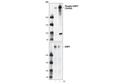
Phospho-53BP1 (Thr543) Antibody is directed at a site that was identified at Cell Signaling Technology (CST) using PhosphoScan®, CST's LC-MS/MS platform for modification site discovery. Phosphorylation at Thr543 was discovered using an ATM/ATR substrate antibody and was shown to be induced by UV treatment. Please visit PhosphoSitePlus®, CST's modification site knowledgebase, at www.phosphosite.org for more information.

Limited Uses

Except as otherwise expressly agreed in a writing signed by a legally authorized representative of CST, the following terms apply to Products provided by CST, its affiliates or its distributors. Any Customer's terms and conditions that are in addition to, or different from, those contained herein, unless separately accepted in writing by a legally authorized representative of CST, are rejected and are of no force or effect.

Products are labeled with For Research Use Only or a similar labeling statement and have not been approved, cleared, or licensed by the FDA or other regulatory foreign or domestic entity, for any purpose. Customer shall not use any Product for any diagnostic or therapeutic purpose, or otherwise in any manner that conflicts with its labeling statement. Products sold or licensed by CST are provided for Customer as the end-user and solely for research and development uses. Any use of Product for diagnostic, prophylactic or therapeutic purposes, or any purchase of Product for resale (alone or as a component) or other commercial purpose, requires a separate license from CST. Customer shall (a) not sell, license, loan, donate or otherwise transfer or make available any Product to any third party, whether alone or in combination with other materials, or use the Products to manufacture any commercial products, (b) not copy, modify, reverse engineer, decompile, disassemble or otherwise attempt to discover the underlying structure or technology of the Products, or use the Products for the purpose of developing any products or services that would compete with CST products or services, (c) not alter or remove from the Products any trademarks, trade names, logos, patent or copyright notices or markings, (d) use the Products solely in accordance with CST Product Terms of Sale and any applicable documentation, and (e) comply with any license, terms of service or similar agreement with respect to any third party products or services used by Customer in connection with the Products.

For Research Use Only. Not for Use in Diagnostic Procedures.
Cell Signaling Technology is a trademark of Cell Signaling Technology, Inc.
All other trademarks are the property of their respective owners. Visit our Trademark Information page.