| Cat. # | Size | Price | Inventory |
|---|---|---|---|
| 9947T | 1 Kit (7 x 20 microliters) |
| Product Includes | Quantity | Applications | Reactivity | MW(kDa) | Isotype |
|---|---|---|---|---|---|
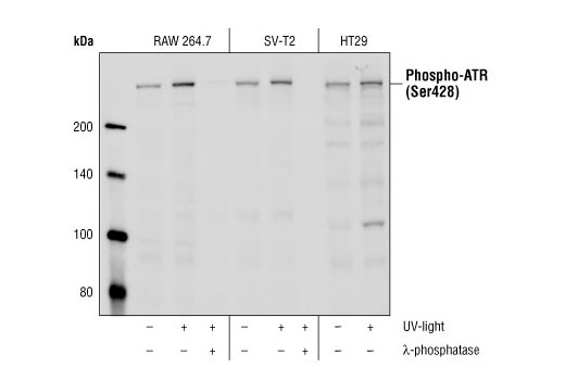
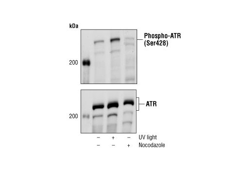
| Phospho-ATR (Ser428) Antibody 2853 | 20 µl |
|
H M R Mk | 300 | Rabbit |
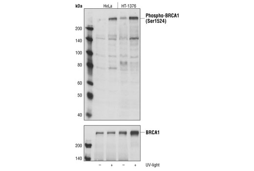
| Phospho-BRCA1 (Ser1524) Antibody 9009 | 20 µl |
|
H | 220 | Rabbit |
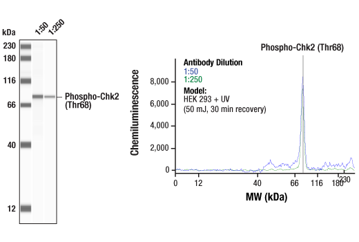
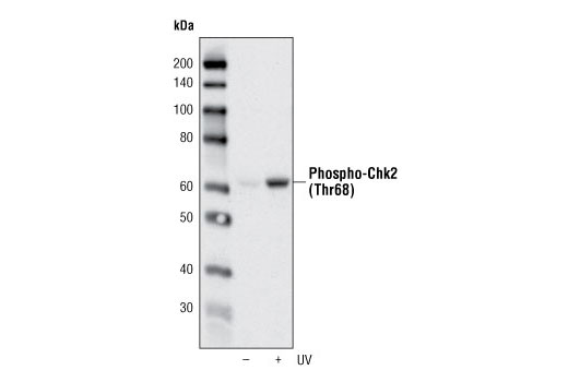
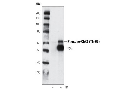
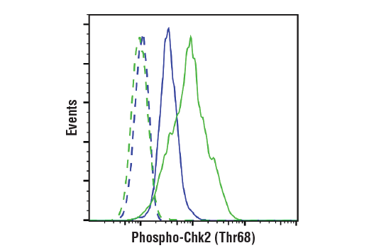
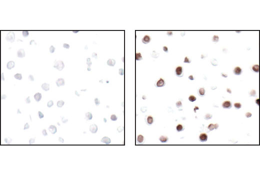
| Phospho-Chk2 (Thr68) (C13C1) Rabbit mAb 2197 | 20 µl |
|
H | 62 | Rabbit IgG |
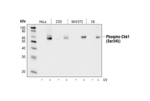
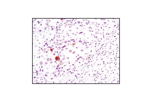
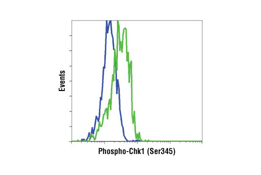
| Phospho-Chk1 (Ser345) (133D3) Rabbit mAb 2348 | 20 µl |
|
H M R Mk | 56 | Rabbit IgG |
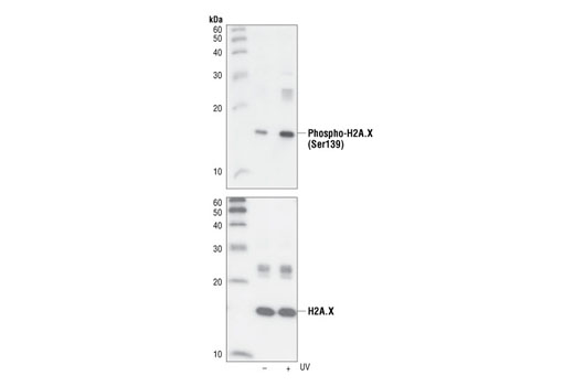
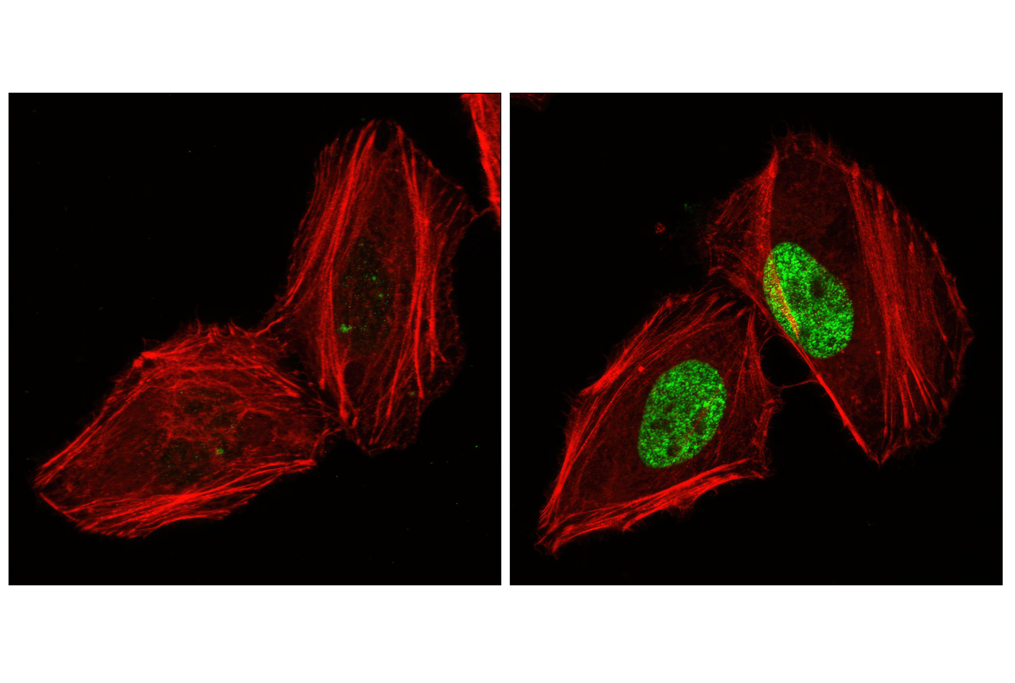
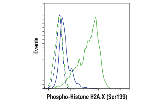
| Phospho-Histone H2A.X (Ser139) (20E3) Rabbit mAb 9718 | 20 µl |
|
H M R Mk | 15 | Rabbit IgG |
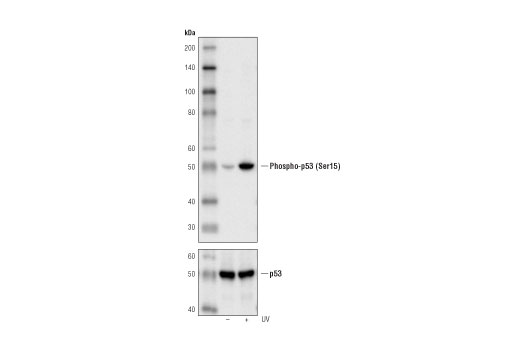
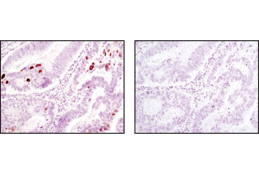
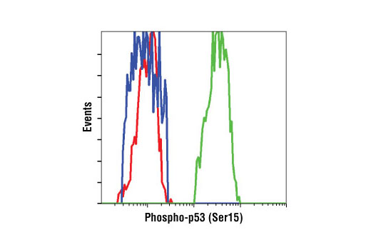
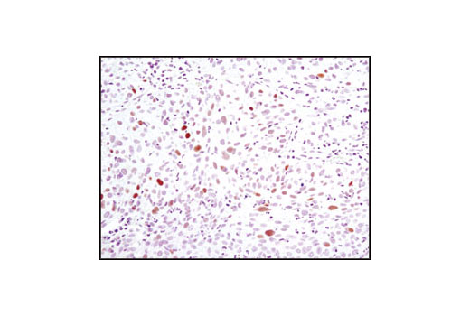
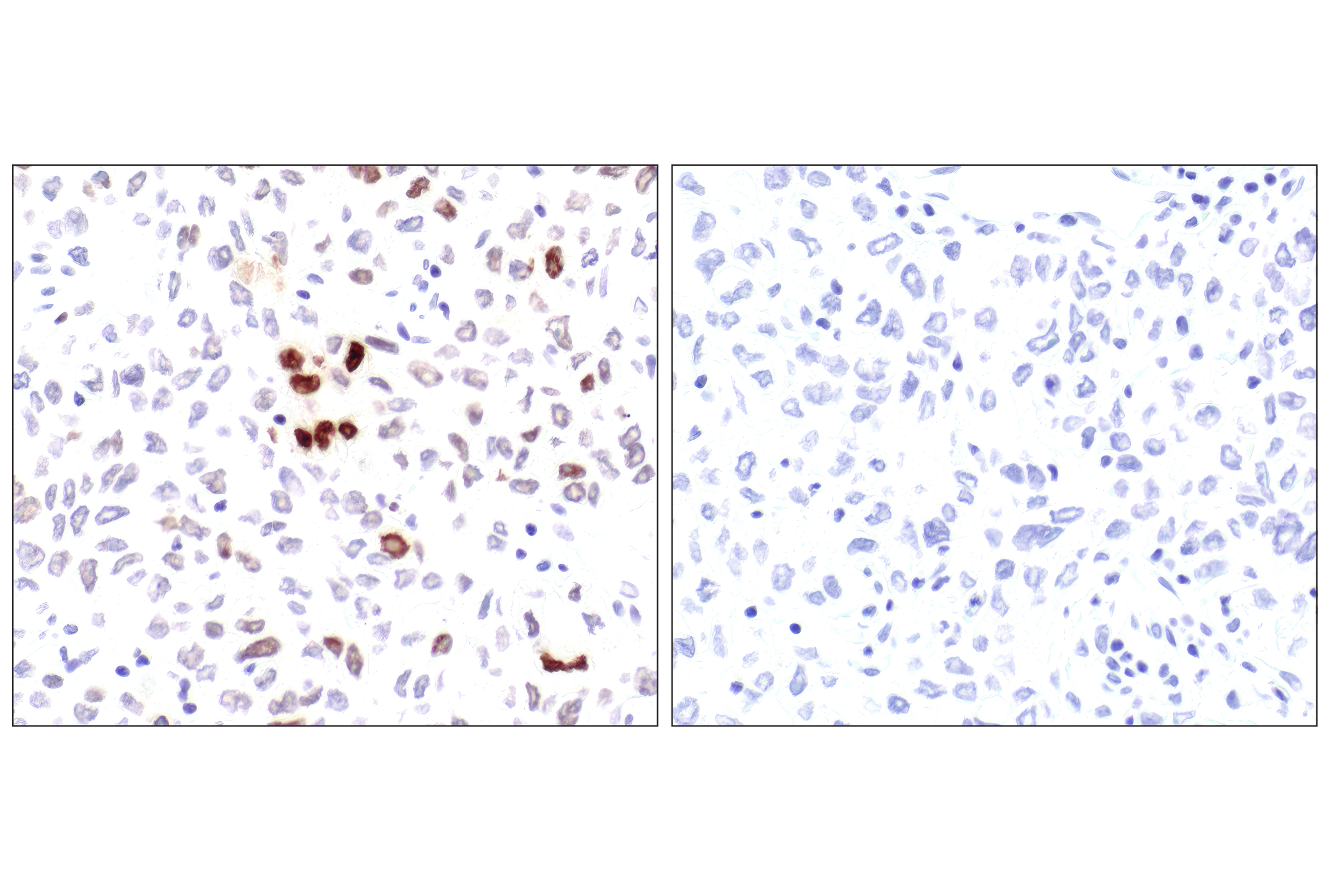
| Phospho-p53 (Ser15) (16G8) Mouse mAb 9286 | 20 µl |
|
H | 53 | Mouse IgG1 |
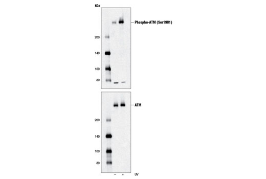
| Phospho-ATM (Ser1981) (D6H9) Rabbit mAb 5883 | 20 µl |
|
H | 350 | Rabbit IgG |
| Anti-rabbit IgG, HRP-linked Antibody 7074 | 100 µl |
|
Goat | ||
| Anti-mouse IgG, HRP-linked Antibody 7076 | 100 µl |
|
Horse |
Product Information
Polyclonal antibodies are produced by immunizing animals with a synthetic peptide and are purified by protein A and peptide affinity chromatography. Monoclonal antibodies are produced by immunizing animals with recombinant human proteins or synthetic peptides.
Ataxia telangiectasia mutated kinase (ATM) and ataxia telangiectasia and Rad3-related kinase (ATR) are PI3 Kinase-related kinase (PIKK) family members that phosphorylate multiple substrates on serine or threonine residues that are followed by a glutamine in response to DNA damage or replication blocks (1-3). p53 is phosphorylated by ATM, ATR and DNA-PK at Ser15. This phosphorylation impairs the ability of MDM2 to bind p53, promoting both the accumulation and activation of p53 in response to DNA damage (4,5). Chk1 and Chk2, downstream protein kinases of ATM/ATR, plays an important role in DNA damage checkpoint control, embryonic development and tumor suppression (6). Chk1 is phosphorylated at Ser280 and Ser296 following DNA damage. The amino-terminal domain of Chk2 contains a series of seven serine or threonine residues, including Thr68, each followed by glutamine (SQ or TQ motif). After DNA damage by ionizing radiation (IR), UV irradiation or hydroxyurea treatment, Thr68 and other sites in this region become phosphorylated by ATM/ATR (7-9). The breast cancer susceptibility proteins BRCA1 and BRCA2 are frequently mutated in cases of hereditary breast and ovarian cancers and have roles in multiple processes related to DNA damage, repair, cell cycle progression, transcription, ubiquitination and apoptosis. Numerous DNA-damage induced phosphorylation sites on BRCA1 have been identified, including serine 1524, and kinases activated in a cell cycle-dependent manner, including Aurora A and CDK2, can also phosphorylate BRCA1. IR, DNA and radiometric-induced DNA damage also results in rapid phosphorylation of the histone H2A family member H2A.X at Ser139 by ATM (10,11). Within minutes following DNA damage, Ser139-phosphorylated H2A.X localizes to sites of DNA damage at subnuclear foci (12).
Except as otherwise expressly agreed in a writing signed by a legally authorized representative of CST, the following terms apply to Products provided by CST, its affiliates or its distributors. Any Customer's terms and conditions that are in addition to, or different from, those contained herein, unless separately accepted in writing by a legally authorized representative of CST, are rejected and are of no force or effect.
Products are labeled with For Research Use Only or a similar labeling statement and have not been approved, cleared, or licensed by the FDA or other regulatory foreign or domestic entity, for any purpose. Customer shall not use any Product for any diagnostic or therapeutic purpose, or otherwise in any manner that conflicts with its labeling statement. Products sold or licensed by CST are provided for Customer as the end-user and solely for research and development uses. Any use of Product for diagnostic, prophylactic or therapeutic purposes, or any purchase of Product for resale (alone or as a component) or other commercial purpose, requires a separate license from CST. Customer shall (a) not sell, license, loan, donate or otherwise transfer or make available any Product to any third party, whether alone or in combination with other materials, or use the Products to manufacture any commercial products, (b) not copy, modify, reverse engineer, decompile, disassemble or otherwise attempt to discover the underlying structure or technology of the Products, or use the Products for the purpose of developing any products or services that would compete with CST products or services, (c) not alter or remove from the Products any trademarks, trade names, logos, patent or copyright notices or markings, (d) use the Products solely in accordance with CST Product Terms of Sale and any applicable documentation, and (e) comply with any license, terms of service or similar agreement with respect to any third party products or services used by Customer in connection with the Products.