查看特别推荐 >>
95235
CD44 (IM7) Rat mAb (Alexa Fluor® 555 Conjugate)
抗体偶联物
单克隆抗体

CD44 (IM7) Rat mAb (Alexa Fluor® 555 Conjugate) #95235

Citations (1)
Filter:
  1. IF
  2. F
Confocal immunofluorescent analysis of Peyer’s patch in mouse small intestine using CD44 (IM7) Rat mAb (Alexa Fluor® 555 Conjugate) (yellow), β-Catenin (L54E2) Mouse mAb (Alexa Fluor® 488 Conjugate) #2849 (green), and CD45 (D3F8Q) Rabbit mAb (Alexa Fluor® 647 Conjugate) #81143 (red). Samples were mounted in ProLong® Gold Antifade Reagent with DAPI #8961 (blue).
Confocal immunofluorescent analysis of C2C12 cells (left, positive) and Neuro-2a cells (right, negative) using CD44 (IM7) Rat mAb (Alexa Fluor® 555 Conjugate) (yellow). Samples were mounted in ProLong® Gold Antifade Reagent with DAPI #8961 (blue).
Flow cytometric analysis of LNCaP cells (blue) and HeLa cells (green) using CD44 (IM7) Rat mAb (Alexa Fluor® 555 Conjugate) (solid lines) or a concentration-matched isotype (dashed lines).
To Purchase # 95235S
Cat. # Size Price Inventory
95235S
100 µl  (50 tests)

Supporting Data

REACTIVITY H M
SENSITIVITY Endogenous
MW (kDa)
Source/Isotype Rat IgG2b kappa

Application Key:

  • WB-Western Blot
  • IP-Immunoprecipitation
  • IHC-Immunohistochemistry
  • ChIP-Chromatin Immunoprecipitation
  • C&R-CUT&RUN
  • C&T-CUT&Tag
  • DB-Dot Blot
  • eCLIP-eCLIP
  • IF-Immunofluorescence
  • F-Flow Cytometry

Species Cross-Reactivity Key:

  • H-Human
  • M-Mouse
  • R-Rat
  • Hm-Hamster
  • Mk-Monkey
  • Vir-Virus
  • Mi-Mink
  • C-Chicken
  • Dm-D. melanogaster
  • X-Xenopus
  • Z-Zebrafish
  • B-Bovine
  • Dg-Dog
  • Pg-Pig
  • Sc-S. cerevisiae
  • Ce-C. elegans
  • Hr-Horse
  • GP-Guinea Pig
  • Rab-Rabbit
  • All-All Species Expected

Product Description

This Cell Signaling Technology antibody is conjugated to Alexa Fluor® 555 fluorescent dye and tested in-house for direct flow cytometric analysis in human cells and immunofluorescent analysis in mouse cells and tissue. This antibody is expected to exhibit the same species cross-reactivity as the unconjugated CD44 (IM7) Rat mAb #39037.

Product Usage Information

Application Dilution
Immunofluorescence (Frozen) 1:200 - 1:800
Immunofluorescence (Immunocytochemistry) 1:200 - 1:800
Flow Cytometry (Fixed/Permeabilized) 1:50
Flow Cytometry (Live) 1:50

Storage

Supplied in PBS (pH 7.2), less than 0.1% sodium azide and 2 mg/ml BSA. Store at 4°C. Do not aliquot the antibody. Protect from light. Do not freeze.

Protocol

PRINT

View >Collapse >

Immunofluorescence (Frozen)

A. Solutions and Reagents

NOTE: Prepare solutions with reverse osmosis deionized (RODI) or equivalent grade water.

  1. 20X Phosphate Buffered Saline (PBS): (9808) To prepare 1L 1X PBS: add 50 ml 20X PBS to 950 ml dH2O, mix. Adjust pH to 8.0.
  2. Formaldehyde: 16%, methanol free, Polysciences, Inc. (cat# 18814), use fresh and store opened vials at 4°C in dark, dilute in 1X PBS for use.
  3. Blocking Buffer (1X PBS / 5% normal goat serum (#5425) / 0.3% Triton™ X-100): To prepare 10 ml: add 0.5 ml normal goat serum and 0.5 ml 20X PBS to 9.0 ml dH2O, mix. While stirring, add 30 µl Triton™ X-100.
  4. Antibody Dilution Buffer: (1X PBS / 1% BSA / 0.3% Triton™ X-100): To prepare 10 ml, add 30 µl Triton™ X-100 to 10 ml 1X PBS. Mix well then add 0.1 g BSA (#9998), mix.
  5. Prolong® Gold AntiFade Reagent (#9071), Prolong® Gold AntiFade Reagent with DAPI (#8961).

B. Specimen Preparation - Frozen/Cryostat Sections (IF-F)

  1. For fixed frozen tissue proceed with Immunostaining (Section C).
  2. For fresh, unfixed frozen tissue, please fix immediately, as follows:
    1. Cover sections with 4% formaldehyde dilute in 1X PBS.

      NOTE: Formaldehyde is toxic, use only in fume hood.

    2. Allow sections to fix for 15 minutes at room temperature.
    3. Aspirate liquid, rinse three times in 1X PBS for 5 minutes each.
    4. Proceed with Immunostaining (Section C).

C. Immunostaining

NOTE: All subsequent incubations should be carried out at room temperature unless otherwise noted in a humid light-tight box or covered dish/plate to prevent drying and fluorochrome fading.

  1. Block specimen in Blocking Buffer for 60 min.
  2. While blocking, prepare primary antibody by diluting as indicated on protocol on product webpage in Antibody Dilution Buffer.
  3. Aspirate blocking solution, apply diluted primary antibody.
  4. Incubate overnight at 4°C.
  5. Rinse three times in 1X PBS for 5 min each.
  6. Coverslip slides with Prolong® Gold Antifade Reagent (#9071) or Prolong® Gold Antifade Reagent with DAPI (#8961).
  7. For best results, allow mountant to cure overnight at room temperature. For long-term storage, store slides flat at 4°C protected from light.

posted November 2006

revised November 2013

Protocol Id: 222

Immunofluorescence (Immunocytochemistry)

A. Solutions and Reagents

NOTE: Prepare solutions with reverse osmosis deionized (RODI) or equivalent grade water.

  1. 20X Phosphate Buffered Saline (PBS): (9808) To prepare 1L 1X PBS: add 50 ml 20X PBS to 950 ml dH2O, mix. Adjust pH to 8.0.
  2. Formaldehyde: 16%, methanol free, Polysciences, Inc. (cat# 18814), use fresh and store opened vials at 4°C in dark, dilute in 1X PBS for use.
  3. Blocking Buffer (1X PBS / 5% normal goat serum (#5425) / 0.3% Triton™ X-100): To prepare 10 ml: add 0.5 ml normal goat serum and 0.5 ml 20X PBS to 9.0 ml dH2O, mix. While stirring, add 30 µl Triton™ X-100.
  4. Antibody Dilution Buffer: (1X PBS / 1% BSA / 0.3% Triton™ X-100): To prepare 10 ml, add 30 µl Triton™ X-100 to 10 ml 1X PBS. Mix well then add 0.1 g BSA (#9998), mix.
  5. Prolong® Gold AntiFade Reagent (#9071), Prolong® Gold AntiFade Reagent with DAPI (#8961).

B. Specimen Preparation - Cultured Cell Lines (IF-IC)

NOTE: Cells should be grown, treated, fixed and stained directly in multi-well plates, chamber slides or on coverslips.

  1. Aspirate liquid, then cover cells to a depth of 2–3 mm with 4% formaldehyde diluted in 1X PBS.

    NOTE: Formaldehyde is toxic, use only in a fume hood.

  2. Allow cells to fix for 15 min at room temperature.
  3. Aspirate fixative, rinse three times in 1X PBS for 5 min each.
  4. Proceed with Immunostaining (Section C).

C. Immunostaining

NOTE: All subsequent incubations should be carried out at room temperature unless otherwise noted in a humid light-tight box or covered dish/plate to prevent drying and fluorochrome fading.

  1. Block specimen in Blocking Buffer for 60 min.
  2. While blocking, prepare primary antibody by diluting as indicated on product webpage in Antibody Dilution Buffer.
  3. Aspirate blocking solution, apply diluted primary antibody.
  4. Incubate overnight at 4°C.
  5. Rinse three times in 1X PBS for 5 min each.
  6. Coverslip slides with Prolong® Gold Antifade Reagent (#9071) or Prolong® Gold Antifade Reagent with DAPI (#8961).
  7. For best results, allow mountant to cure overnight at room temperature. For long-term storage, store slides flat at 4°C protected from light.

posted November 2006

revised November 2013

Protocol Id: 182

Flow Cytometry, Methanol Permeabilization Protocol for Directly Conjugated Antibodies

A. Solutions and Reagents

All reagents required for this protocol may be efficiently purchased together in our Intracellular Flow Cytometry Kit (Methanol) #13593, or individually using the catalog numbers listed below.

NOTE: Prepare solutions with reverse osmosis deionized (RODI) or equivalent grade water.

  1. 1X Phosphate Buffered Saline (PBS): To prepare 1 L 1X PBS: add 100 ml 10X PBS (#12528) to 900 ml water mix.
  2. 4% Formaldehyde, Methanol-Free (#47746)
  3. 100% Methanol (#13604): Chill before use
  4. Antibody Dilution Buffer: Purchase ready-to-use Flow Cytometry Antibody Dilution Buffer (#13616), or prepare a 0.5% BSA PBS buffer by dissolving 0.5 g Bovine Serum Albumin (BSA) (#9998) in 100 ml 1X PBS. Store at 4°C.

NOTE: When including fluorescent cellular dyes in your experiment (including viability dyes, DNA dyes, etc.), please refer to the dye product page for the recommended protocol. Visit www.cellsignal.com for a full listing of cellular dyes validated for use in flow cytometry.

B. Fixation

NOTE: Adherent cells or tissue should be dissociated and in single-cell suspension prior to fixation.

NOTE: Optimal centrifugation conditions will vary depending upon cell type and reagent volume. Generally, 150-300g for 1-5 minutes will be sufficient to pellet the cells.

NOTE: If using whole blood, lyse red blood cells and wash by centrifugation prior to fixation.

NOTE: Antibodies targeting CD markers or other extracellular proteins may be added prior to fixation if the epitope is disrupted by formaldehyde and/or methanol. The antibodies will remain bound to the target of interest during the fixation and permeabilization process. However, note that some fluorophores (including PE and APC) are damaged by methanol and thus should not be added prior to permeabilization. Conduct a small-scale experiment if you are unsure.

  1. Pellet cells by centrifugation and remove supernatant.
  2. Resuspend cells in approximately 100 µl 4% formaldehyde per 1 million cells. Mix well to dissociate pellet and prevent cross-linking of individual cells.
  3. Fix for 15 min at room temperature (20-25°C).
  4. Wash by centrifugation with excess 1X PBS. Discard supernatant in appropriate waste container. Resuspend cells in 0.5-1 ml 1X PBS. Proceed to Permeabilization step.
    1. Alternatively, cells may be stored overnight at 4°C in 1X PBS.

C. Permeabilization

  1. Permeabilize cells by adding ice-cold 100% methanol slowly to pre-chilled cells, while gently vortexing, to a final concentration of 90% methanol.
  2. Permeabilize for a minimum of 10 min on ice.
  3. Proceed with immunostaining (Section D) or store cells at -20°C in 90% methanol.

D. Immunostaining

NOTE: Count cells using a hemocytometer or alternative method.

  1. Aliquot desired number of cells into tubes or wells. (Generally, 5x10⁵ to 1x10⁶ cells per assay.)
  2. Wash cells by centrifugation in excess 1X PBS to remove methanol. Discard supernatant in appropriate waste container. Repeat if necessary.
  3. Resuspend cells in 100 µl of diluted primary antibody, prepared in Antibody Dilution Buffer at a recommended dilution or as determined via titration.
  4. Incubate for 1 hr at room temperature. Protect from light.
  5. Wash by centrifugation in Antibody Dilution Buffer or 1X PBS. Discard supernatant. Repeat.
  6. Resuspend cells in 200-500 µl of 1X PBS and analyze on flow cytometer.

posted July 2009

revised June 2020

实验步骤编号:407

流式细胞术,针对直接结合抗体的活细胞实验步骤

A. 溶液与试剂

:使用反渗透去离子水 (RODI) 或同等级别的水配制溶液。

  1. 1 X 磷酸盐缓冲生理盐水 (PBS):要配制 1 L 1X PBS:将 100 ml 10X PBS (#12528) 添加到 900 ml 水,混合。
  2. 抗体稀释缓冲液:购买即用型流式细胞术抗体稀释缓冲液 (#13616),或在 100 ml 1x PBS 中溶解 0.5 g 牛血清白蛋白 (BSA) (#9998) 来配制 0.5 % BSA PBS 缓冲液。4°C 保存。

:在您的实验中加入荧光细胞染料(包括活力指示染料、DNA 染料等)时,请参考染料产品网页,了解建议的实验步骤。访问 www.cellsignal.com,了解经验证用于流式细胞术的细胞染料完整列表。

B. 免疫染色

:使用血细胞计数器或备选方法计数细胞。

:如果使用全血,则需裂解红血细胞,并在免疫染色之前通过离心分离洗涤。

:人 Fc 受体与兔 IgG 发生交叉反应。当感兴趣的细胞表达高水平的 Fc 受体蛋白(例如,巨噬细胞/单核细胞谱系)时,在用兔抗体进行免疫染色之前,用人 Fc 块预孵育活细胞。

:最佳离心条件会根据细胞类型和试剂容量变动。一般,1-5 分钟 150-300g 将足以使细胞沉淀下来。

  1. 将所需数目的细胞分装入试管或小孔中。(通常,每次测定 5 x 10⁵ 至 1 x 10⁶ 个细胞。)
  2. 通过离心使细胞沉淀下来,移除上清液。
  3. 在 100 µl 稀释的一抗中重悬细胞,这种一抗按建议的稀释度或如通过滴定所确定那样以抗体稀释缓冲液配制。
  4. 在冰上孵育 30 分钟至 1 小时。避光。
  5. 用抗体稀释缓冲液进行离心洗涤。弃去上清液。重复。
  6. 在 200-500 μl 抗体稀释缓冲液中重悬细胞后用流式细胞分析仪进行分析。

发布​时间 2017 年 6 月

修订时间 2022 年 1 月

实验步骤编号:1504

特异性/灵敏度

CD44 (IM7) Rat mAb (Alexa Fluor® 555 Conjugate) 可检测内源水平的 CD44 总蛋白。该抗体可检测细胞外结构域内的表位,并且预计可检测所有 CD44 同工型。除小鼠反应性外,流式细胞分析显示该克隆还对人样品呈阳性。

物种反应性:

人, 小鼠

来源/纯化

通过亲和色谱从组织培养上清液纯化这种单克隆抗体。

背景

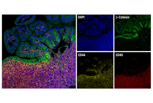
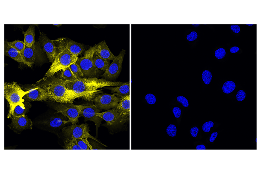
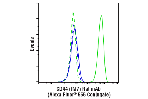
CD44 是 I 型跨膜糖蛋白,可通过对透明质酸 (HA) 的亲和力并可能通过胞外基质 (ECM) 的其他部分,介导细胞-细胞相互作用和细胞-基质相互作用。CD44 呈高度多态性,具有多种选择性剪切变体, 经受广泛的翻译后修饰 (1,2)。CD44 的表面水平升高是 T 细胞活化的特征,并且该蛋白质的表达在炎症反应期间上调。研究已经证实, CD44 和 HER2 之间的相互作用与卵巢癌细胞生长的增长相关 (1-3)。CD44 与埃兹蛋白、根蛋白和膜突蛋白 (ERM) 相互作用,使肌动蛋白细胞骨架与质膜和 ECM 连接 (4-6)。在静息细胞中,CD44 在 Ser325 处组成型磷酸化。PKC 的激活导致 Ser291 磷酸化、Ser325 去磷酸化、埃兹蛋白从 CD44 解离和定向运动性 (4)。

有限使用

除非如以 CST 合法授权代表签署的书面形式另行明确同意,否则以下条款适用于 CST、其附属公司或其分销商提供的产品。除非 CST 合法授权代表以书面形式单独接受,否则任何附加于或异于此处所载条款和条件的客户条款和条件均被拒绝且无效。

产品用“仅供研究使用”或类似标示声明标示,并且尚未经 FDA 或其他国外或国内监管实体出于任何目的批准、准许或许可。客户不得出于任何诊断或治疗目的或以任何与产品标示声明相冲突的方式使用任何产品。CST 销售或许可的产品提供给作为最终用户的客户,且仅用于研究和开发用途。出于诊断、预防或治疗目的任何产品使用或出于转售(单独或作为成分)或其他商业目的的任何产品购买都要求来自 CST 的单独许可。客户 (a) 不得向任何第三方出售、许可、出借、捐赠或另行转让或提供任何本公司产品,无论单独或联合其他材料方式,或使用本公司产品制造任何商业产品,(b) 不得复制、修改、逆向工程、反编译、反汇编或另行尝试发现本公司产品的底层结构或技术,或出于开发与 CST 产品或服务竞争的任何产品或服务的目的使用本公司产品,(c) 不得从本公司产品改变或移除任何商标、商品名称、徽标、专利或版权声明或标记,(d) 仅应根据 CST 产品销售条款和任何适用文档使用本公司产品,以及 (e) 应就客户联系本公司产品所用的任何第三方产品或服务而言遵守任何许可、服务条款或类似协议。

仅供研究使用。不得用于诊断流程。
Cell Signaling Technology 是 Cell Signaling Technology, Inc. 的商标。
本产品依据源于 Life Technologies Corporation 的知识产权许可提供。本产品的转让条件为买方仅在买方开展的研究中使用所购买的产品,不包括合同研究或任何服务收费研究,并且买方不得 (1) 将本产品或其组分用于以下方面:(a) 用于诊断、诊疗或预防目的;(b) 用于检测、分析或筛选服务或获得信息,以按检测收取报酬;或 (c) 用于制造或质量保证或质量控制,和/或 (2) 销售或转让本产品或其组分用于转售,无论是否转售供研究使用。如需了解有关购买本品用于除上述目的之外目的的许可信息,请联系 Life Technologies Corporation(地址:5791Van Allen Way, Carlsbad, CA 92008 USA)或 [受保护的电子邮件]
所有其他商标均属各自所有者专有。访问我们的商标信息页面。